Breadcrumb
- Home
- Research
Research
Our research focuses on using medical imaging to understand the normal and diseased lung. This work involves novel image acquisition methods, image processing and visualization, and data analysis, machine learning, and modeling. Our current projects are in five main areas, summarized below. A more complete list of publications is available on our lab publications page or on Google Scholar.
- Pulmonary Image Analysis: We have developed a large collection of robust and validated image analysis algorithms and tools to facilitate quantitative lung imaging, including tools to identify the lungs, lobes, airways, and blood vessels in CT images, algorithms for airway caliber measurement and airway tree analysis, and image registration methods for detecting changes in longitudinal studies. Many of these algorithms have been commercialized by VIDA Diagnostics, Inc., a University of Iowa spin-off company. Some of the recent papers in this area include:
- Gerard SE, Herrmann J, Xin Y, Martin KT, Rezoagli E, Ippolito D, Bellani G, Cereda M, Guo J, Hoffman EA, Kaczka DW, Reinhardt JM. CT image segmentation for inflamed and fibrotic lungs using a multi-resolution convolutional neural network. Sci Rep. 2021 Jan 14;11(1):1455. PubMed Central PMCID: PMC7809065.
- Gerard SE, Herrmann J, Kaczka DW, Musch G, Fernandez-Bustamante A, Reinhardt JM. Multi-resolution convolutional neural networks for fully automated segmentation of acutely injured lungs in multiple species. Med Image Anal. 2020 Feb;60:101592. PubMed Central PMCID: PMC6980773.
- Gerard SE, Patton TJ, Christensen GE, Bayouth JE, Reinhardt JM. FissureNet: A Deep Learning Approach For Pulmonary Fissure Detection in CT Images. IEEE Trans Med Imaging. 2019 Jan;38(1):156-166. PubMed Central PMCID: PMC6318012.
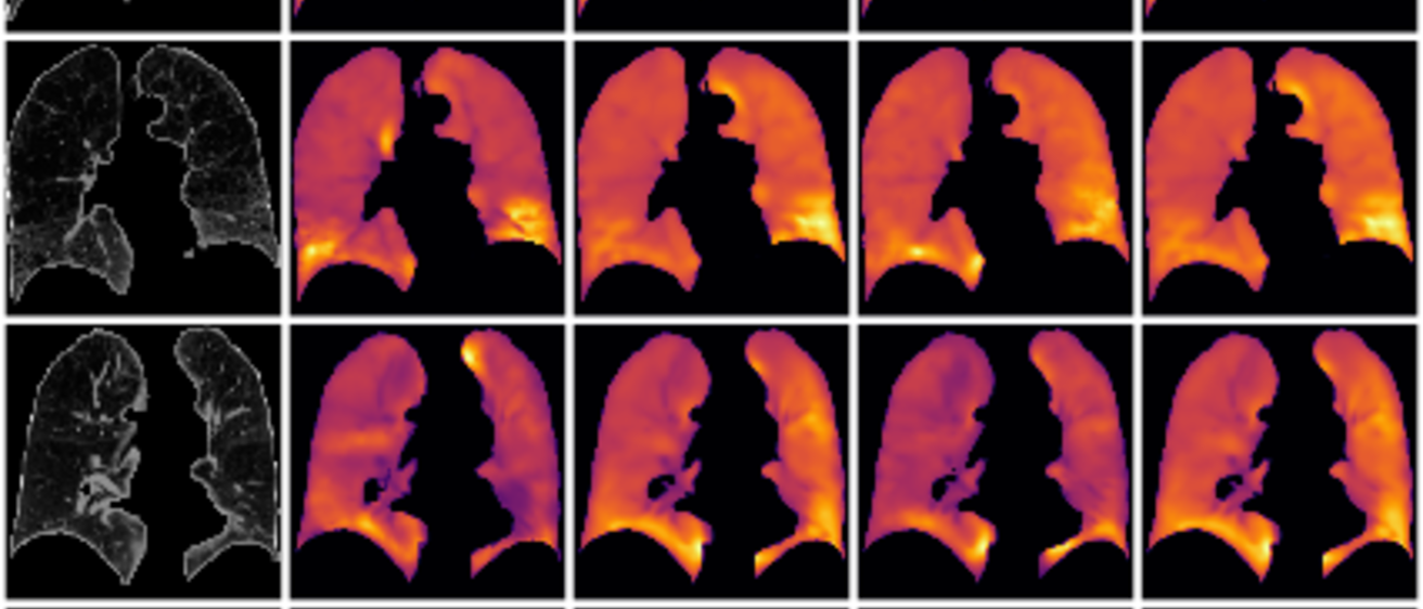
- Measuring Regional Lung Function: Our lab has been actively developing the use of 3D and 4D X-ray CT as a modality to provide robust and repeatable estimates of regional pulmonary function. Using respiratory gated imaging and image registration, we calculate the local tissue expansion and contraction across volume changes. We have compared these measures to xenon CT measures of ventilation and to spirometry to establish physiologic significance. Some papers describing this work include:
- Patton TJ, Gerard SE, Shao W, Christensen GE, Reinhardt JM, Bayouth JE. Quantifying ventilation change due to radiation therapy using 4DCT Jacobian calculations. Med Phys. 2018 Oct;45(10):4483-4492. PubMed Central PMCID: PMC6220845.
- Ding K, Cao K, Fuld MK, Du K, Christensen GE, Hoffman EA, Reinhardt JM. Comparison of image registration based measures of regional lung ventilation from dynamic spiral CT with Xe-CT. Med Phys. 2012 Aug;39(8):5084-98. PubMed Central PMCID: PMC3416881.
- Ding K, Bayouth JE, Buatti JM, Christensen GE, Reinhardt JM. 4DCT-based measurement of changes in pulmonary function following a course of radiation therapy. Med Phys. 2010 Mar;37(3):1261-72. PubMed Central PMCID: PMC2842288.
- Reinhardt JM, Ding K, Cao K, Christensen GE, Hoffman EA, Bodas SV. Registration-based estimates of local lung tissue expansion compared to xenon CT measures of specific ventilation. Med Image Anal. 2008 Dec;12(6):752-63. PubMed Central PMCID: PMC2692217.
- Hu S, Hoffman EA, Reinhardt JM. Automatic lung segmentation for accurate quantitation of volumetric X-ray CT images. IEEE Trans Med Imaging. 2001 Jun;20(6):490-8. PubMed PMID: 11437109.
- Lung Mechanics in COPD: My group has studied regional lung mechanics in normals and subject with COPD and related disorders. Our method is based on respiratory-gated image acquisition and 3D image registration to track local tissue deformation. We have described lobar deformation and sliding across the respiratory cycle, introduced a quantitative measure to characterize sliding on the lung borders, and applied these methods within a machine learning framework to look at differences in biomechanics between normals and subjects with COPD. Recent papers in this area include:
- Bhatt SP, Bodduluri S, Hoffman EA, Newell JD Jr, Sieren JC, Dransfield MT, Reinhardt JM. Computed Tomography Measure of Lung at Risk and Lung Function Decline in Chronic Obstructive Pulmonary Disease. Am J Respir Crit Care Med. 2017 Sep 1;196(5):569-576. PubMed Central PMCID: PMC5620667.
- Bodduluri S, Bhatt SP, Hoffman EA, Newell JD Jr, Martinez CH, Dransfield MT, Han MK, Reinhardt JM. Biomechanical CT metrics are associated with patient outcomes in COPD. Thorax. 2017 May;72(5):409-414. PubMed Central PMCID: PMC5526353.
- Bhatt SP, Bodduluri S, Newell JD, Hoffman EA, Sieren JC, Han MK, Dransfield MT, Reinhardt JM. CT-derived Biomechanical Metrics Improve Agreement Between Spirometry and Emphysema. Acad Radiol. 2016 Oct;23(10):1255-63. PubMed Central PMCID: PMC5026854.
- Bodduluri S, Newell JD Jr, Hoffman EA, Reinhardt JM. Registration-based lung mechanical analysis of chronic obstructive pulmonary disease (COPD) using a supervised machine learning framework. Acad Radiol. 2013 May;20(5):527-36. PubMed Central PMCID: PMC3644222.
- Studying Normal Lung Biomechanics: Using respiratory-gated CT imaging, we have studied the biomechanics of the normal lung during respiration by calculating lung tissue strain, anisotropic deformation index (a measure of anisotropy), and Jacobian determinant (a measure of local volume change). Some of our recent work in this area is:
- Amelon R, Cao K, Ding K, Christensen GE, Reinhardt JM, Raghavan ML. Three-dimensional characterization of regional lung deformation. J Biomech. 2011 Sep 2;44(13):2489-95. PubMed Central PMCID: PMC3443473.
- Amelon RE, Cao K, Reinhardt JM, Christensen GE, Raghavan ML. A measure for characterizing sliding on lung boundaries. Ann Biomed Eng. 2014 Mar;42(3):642-50. PubMed Central PMCID: PMC3943475.
- Cao K, Christensen GE, Ding K, Du K, Raghavan ML, Amelon RE, Baker KM, Hoffman EA, Reinhardt JM. Tracking regional tissue volume and function change in lung using image registration. Int J Biomed Imaging. 2012;2012:956248. PubMed Central PMCID: PMC3483832.
- Ding K, Yin Y, Cao K, Christensen GE, Lin CL, Hoffman EA, Reinhardt JM. Evaluation of lobar biomechanics during respiration using image registration. Med Image Comput Comput Assist Interv. 2009;12(Pt 1):739-46. PubMed Central PMCID: PMC2913717.
- Functional Avoidance Strategies for Radiation Therapy: We have developed models of radiation-induced lung tissue damage and created strategies for guiding radiation therapy using CT-based lung function analysis. Our recent papers for this project include:
- Wallat EM, Flakus MJ, Wuschner AE, Shao W, Christensen GE, Reinhardt JM, Baschnagel AM, Bayouth JE. Modeling the impact of out-of-phase ventilation on normal lung tissue response to radiation dose. Med Phys. 2020 Jul;47(7):3233-3242. PubMed PMID: 32187683.
- Kipritidis J, Tahir BA, Cazoulat G, Hofman MS, Siva S, Callahan J, Hardcastle N, Yamamoto T, Christensen GE, Reinhardt JM, Kadoya N, Patton TJ, Gerard SE, Duarte I, Archibald-Heeren B, Byrne M, Sims R, Ramsay S, Booth JT, Eslick E, Hegi-Johnson F, Woodruff HC, Ireland RH, Wild JM, Cai J, Bayouth JE, Brock K, Keall PJ. The VAMPIRE challenge: A multi-institutional validation study of CT ventilation imaging. Med Phys. 2019 Mar;46(3):1198-1217. PubMed Central PMCID: PMC6605778.
- Patton TJ, Gerard SE, Shao W, Christensen GE, Reinhardt JM, Bayouth JE. Quantifying ventilation change due to radiation therapy using 4DCT Jacobian calculations. Med Phys. 2018 Oct;45(10):4483-4492. PubMed Central PMCID: PMC6220845.
- Du K, Reinhardt JM, Christensen GE, Ding K, Bayouth JE. Respiratory effort correction strategies to improve the reproducibility of lung expansion measurements. Med Phys. 2013 Dec;40(12):123504. PubMed Central PMCID: PMC3843762.