A new paper entitled "Direct estimation of regional lung volume change from paired and single CT images using residual regression neural network" was just published in Medical Physics.
Background
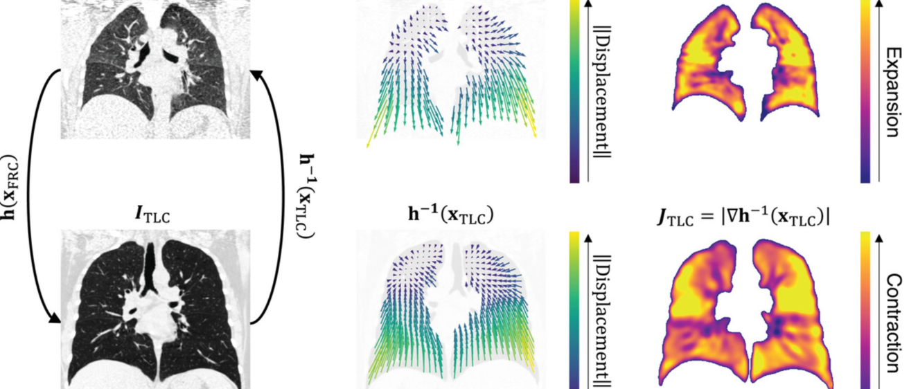
Chest computed tomography (CT) enables characterization of pulmonary diseases by producing high-resolution and high-contrast images of the intricate lung structures. Deformable image registration is used to align chest CT scans at different lung volumes, yielding estimates of local tissue expansion and contraction.
Purpose
We investigated the utility of deep generative models for directly predicting local tissue volume change from lung CT images, bypassing computationally expensive iterative image registration and providing a method that can be utilized in scenarios where either one or two CT scans are available.
Methods
A residual regression convolutional neural network, called Reg3DNet+, is proposed for directly regressing high-resolution images of local tissue volume change (i.e., Jacobian) from CT images. Image registration was performed between lung volumes at total lung capacity (TLC) and functional residual capacity (FRC) using a tissue mass- and structure-preserving registration algorithm. The Jacobian image was calculated from the registration-derived displacement field and used as the ground truth for local tissue volume change. Four separate Reg3DNet+ models were trained to predict Jacobian images using a multifactorial study design to compare the effects of network input (i.e., single image vs. paired images) and output space (i.e., FRC vs. TLC). The models were trained and evaluated on image datasets from the COPDGene study. Models were evaluated against the registration-derived Jacobian images using local, regional, and global evaluation metrics.
Results
Statistical analysis revealed that both factors – network input and output space – were significant determinants for change in evaluation metrics. Paired-input models performed better than single-input models, and model performance was better in the output space of FRC rather than TLC. Mean structural similarity index for paired-input models was 0.959 and 0.956 for FRC and TLC output spaces, respectively, and for single-input models was 0.951 and 0.937. Global evaluation metrics demonstrated correlation between registration-derived Jacobian mean and predicted Jacobian mean: coefficient of determination (r2) for paired-input models was 0.974 and 0.938 for FRC and TLC output spaces, respectively, and for single-input models was 0.598 and 0.346. After correcting for effort, registration-derived lobar volume change was strongly correlated with the predicted lobar volume change: for paired-input models r2 was 0.899 for both FRC and TLC output spaces, and for single-input models r2 was 0.803 and 0.862, respectively.
Conclusions
Convolutional neural networks can be used to directly predict local tissue mechanics, eliminating the need for computationally expensive image registration. Networks that use paired CT images acquired at TLC and FRC allow for more accurate prediction of local tissue expansion compared to networks that use a single image. Networks that only require a single input image still show promising results, particularly after correcting for effort, and allow for local tissue expansion estimation in cases where multiple CT scans are not available. For single-input networks, the FRC image is more predictive of local tissue volume change compared to the TLC image.